Medical image processing
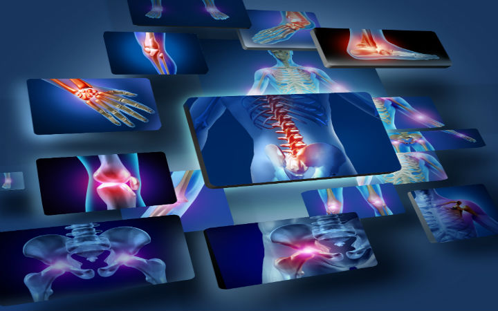
Medical image processing is a specialized field that focuses on analyzing and interpreting medical images to enhance diagnosis, treatment planning, and patient monitoring. Using advanced computational techniques, medical image processing aims to extract meaningful information from various imaging modalities, including X-ray, MRI, CT, ultrasound, and PET scans. This field combines elements of computer vision, image analysis, and machine learning to aid healthcare professionals in accurately identifying abnormalities, monitoring disease progression, and evaluating treatment outcomes.
At the heart of medical image processing lies the challenge of translating complex visual data into actionable insights. Techniques such as image segmentation, feature extraction, and image reconstruction allow for precise delineation of anatomical structures, identification of pathological changes, and quantitative analysis of tissue characteristics. For instance, segmentation algorithms can help isolate organs, tumors, or lesions, providing crucial information for oncologists, radiologists, and surgeons. Feature extraction, on the other hand, focuses on identifying specific attributes within an image—such as texture, shape, or intensity—that are indicative of disease, which can be especially helpful in early detection and personalized treatment.
Machine learning, particularly deep learning, plays a significant role in medical image processing by enabling the automation and enhancement of diagnostic procedures. Convolutional neural networks (CNNs) are widely used to classify medical images, detect anomalies, and even predict disease risk with remarkable accuracy. These models are trained on large datasets to recognize patterns that may not be immediately visible to the human eye, helping to improve diagnostic speed and accuracy. Additionally, AI-powered image processing tools are being developed to assist in predicting treatment responses, optimizing radiotherapy plans, and tracking patient recovery over time.
Beyond diagnostics, medical image processing contributes to advancements in minimally invasive surgeries and robotics. Image-guided surgery, for example, uses real-time imaging to assist surgeons in navigating complex anatomical structures with precision, reducing the risks associated with traditional surgical methods. Furthermore, 3D reconstruction and visualization allow for more effective preoperative planning and education, giving both clinicians and patients a better understanding of medical procedures and expected outcomes.
Medical image processing also addresses the challenges of image quality and standardization, which are essential for reliable interpretation. Techniques such as noise reduction, contrast enhancement, and resolution improvement ensure that images are clear and consistent across different machines and clinical settings. This is particularly important in telemedicine, where high-quality imaging allows remote specialists to make accurate assessments.
In the future, medical image processing is expected to advance with the integration of real-time analytics, cloud computing, and enhanced AI models. These developments hold the potential to streamline workflows in clinical settings, provide rapid and precise diagnostic insights, and open new possibilities in personalized and predictive medicine. As medical imaging technology continues to evolve, medical image processing stands at the forefront, transforming healthcare by improving patient outcomes and enhancing our understanding of the human body through powerful visualization and analysis tools.

2025
, (2025). IRAU-Net: Inception Residual Attention U-Net in Adversarial Network for Cardiac MRI Segmentation. IEEE Journal of Selected Topics in Signal Processing, (), . DOI: https://doi.org/10.1109/JSTSP.2024.3523233


